Finance cosmetic procedures with as little as $0 down! Click here to apply. Or, learn more here.
Get accurate, safer dermal filler treatments and corrections with our ultrasound device

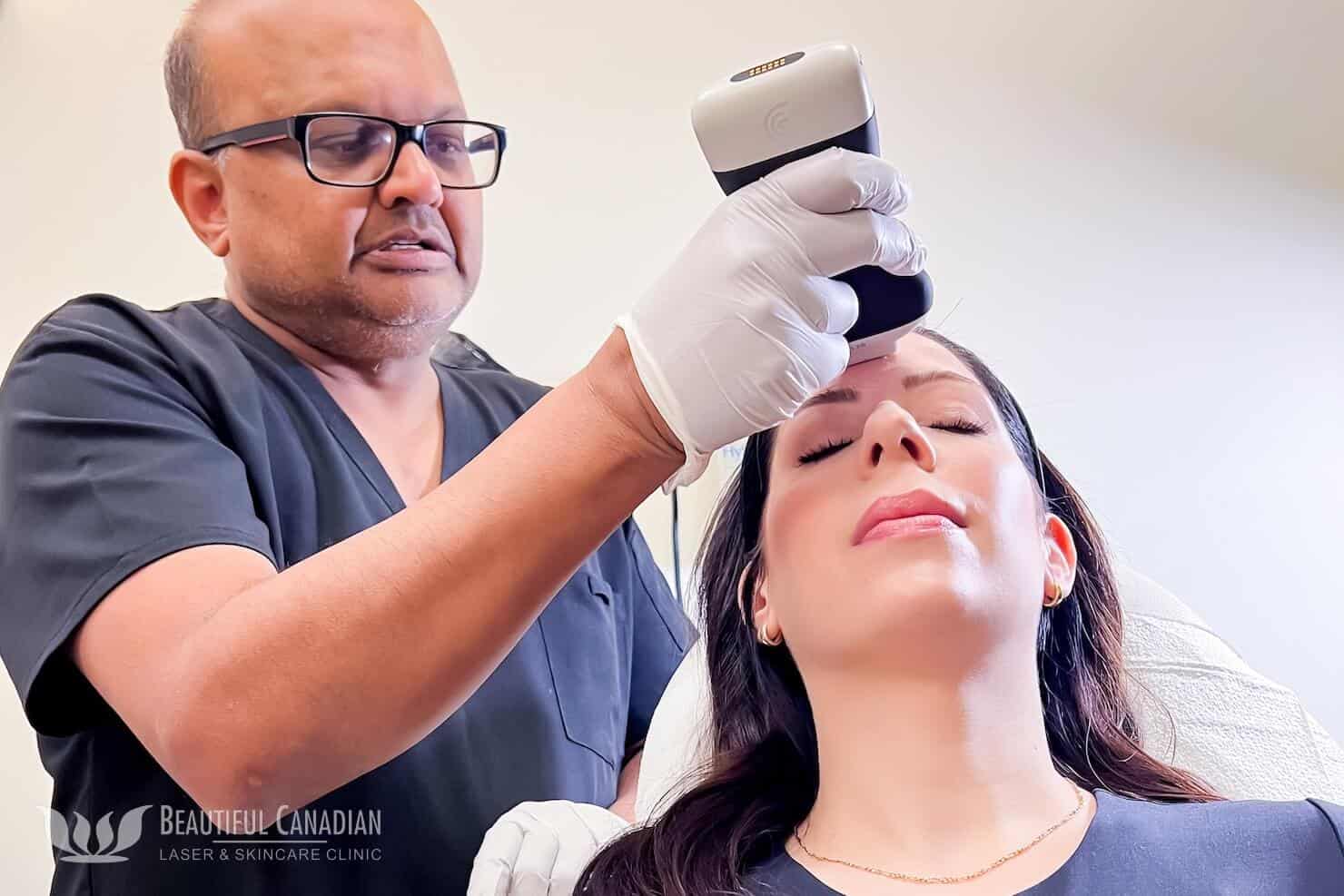
At our clinic, Surrey dermal fillers are safer and more accurate thanks to the Clarius ultrasound system. The cost of an Ultrasound for Filler is $125, and your Consultation Fee will be applied toward this service!
What is the Clarius ultrasound device?
The Clarius ultrasound device is a Canadian-made tool used by medical doctors in an array of applications, including aesthetics. It outputs high definition (HD) images on a phone or tablet, via a scanner that comes into contact with your skin.
Ultrasound helps medical practitioners see under your skin, without opening it.
Based on the settings selected by a medical practitioner, quality ultrasound software can highlight specific bodily structures or implanted materials. The Clarius device comes with a software made specifically for use in medical aesthetics.
Why do we use ultrasound during your dermal filler treatments?
There are two main reasons why we use ultrasound imaging during your dermal filler treatments:
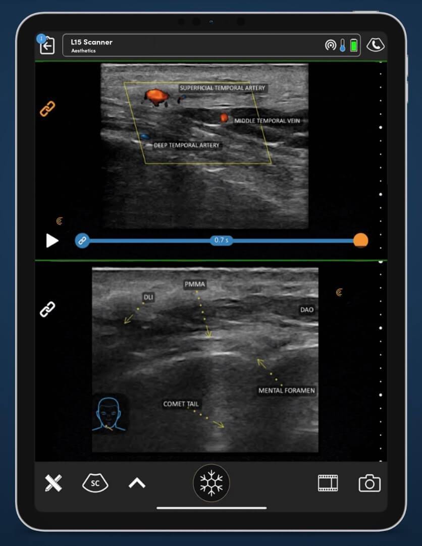
Avoiding veins and arteries for your safety

Ultrasound imaging allows us to avoid vascular structures in your dermis while injecting dermal fillers. This is important because vascular occlusions (i.e. blood vessel blockages) are a known side effect of dermal fillers. Albeit, this can occur largely due to technique.
Live ultrasound imaging shows us clearly where your veins, arteries and other anatomical structures are.
Of course, the study of facial anatomy is a critical part of medical aesthetics training. This knowledge informs our injections, as always. However, for patients who have had prior surgery, veins and arteries may have moved. This makes dermal filler placement a tad more complicated.
Our skilled injectors have previously used the cannula method to provide safe, accurate injections (which is advanced, since this method is not always used by all injectors!).
But now, doctors and nurses can use cannulas alongside ultrasound.
The added visual guidance makes the treatments even more safe and accurate. In fact, the live imaging can show the cannula’s placement under the skin while fillers are being injected!
Providing symmetrical, better results where past fillers have been injected

Sometimes, we see patients who have had previous dermal filler injected by external providers. However, they are not sure where, or what kind of dermal fillers were used.
Knowing this information is important because different dermal fillers behave differently under the skin.
For example, hyaluronic acid (HA) fillers like Juvéderm® act as a gel that attracts water. This gives your skin an immediate ‘boost’, but fades over time.
On the other hand, there are fillers like Sculptra®, which uses poly-L-lactic acid (PLLA). This substance kick starts the body’s own collagen mechanisms. Then, over months, more collagen is produced in the dermis. The added collagen lifts the skin and makes it naturally ‘plump.’
And so on and so forth with other types of fillers.
Hyaluronic acid is dissolvable with hyaluronidase, whereas fillers made of other materials are not.
So, when deciding where, how much, and the type of filler to place at injection points, it helps tremendously to know what other materials are already in the dermis. An injector can then pre-plan for future collagen growth, loss of volume, or potential migrations of filler.
They can also more precisely inject around other fillers, rather than through them.
How can ultrasound help correct or remove dermal filler treatments?

Ultrasound imaging is also useful in cases where someone doesn’t like their dermal filler treatment, or their dermal filler has migrated from its original placement.
If the dermal filler is made of hyaluronic acid (HA), then hyaluronidase can be used to dissolve it.
However, in order for hyaluronidase to be effective, it needs to be injected precisely where the HA filler lies. Otherwise, it won’t work as well.
The handheld ultrasound device at our clinic allows us to find existing dermal filler deep under your skin. This means we don’t have to rely on touch alone to find it. We can see it visually on a screen. Then, we can dissolve it with hyaluronidase.
Once dissolved, HA dermal filler should dissipate, and your face should go back to the way it was before you had filler injected.
Choose the most advanced dermal filler treatments available for your peace of mind

Dermal filler is popular, but it’s not the same as buying cosmetics at the drugstore. It’s a medical procedure.
By choosing skilled injectors who use the latest technology available, you can protect your investment in dermal fillers and avoid potential side effects.
At our clinic, we aim to ‘stay ahead of the game’ by learning and acquiring new technological tools that make our services better.
For you, ultrasound imaging with dermal filler treatments means better, more accurate results. It also means safety and peace of mind.
